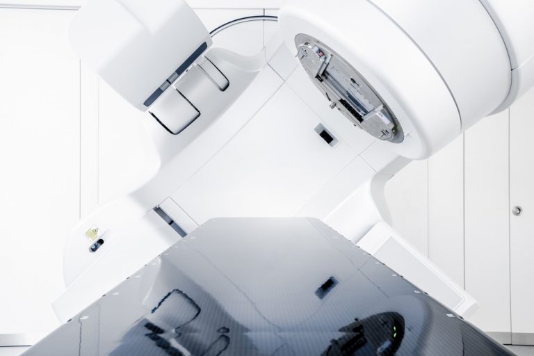
Radiation is a form of cancer treatment that uses externally generated energy rays to target and kill cancer cells. This therapy works by destroying the DNA in cells it targets and leads to cancer cell death. Many forms of cancer can be treated with radiation especially prostate cancer.
Radiation is generally delivered from a machine outside the body (external beam radiation) but can also be given by placing radioactive implants inside the body (brachytherapy). External beam radiation (EBRT) is generally delivered in the form of photon beams. A photon is a form of electromagnetic energy that can be thought of as a bundle of energy. Photons are created in a linear accelerator and then delivered to the patient.Unfortunately, rays can also affect healthy, non-cancerous cells and this can lead to side effects from radiation treatment. It is extremely important to be able to precisely target cancer cells while minimizing collateral damage to the nearby healthy cells.
Recent advances in technology have allowed physicians to develop a sophisticated radiation technique called: IMRT-IGRT, which stands for Intensity Modulated Radiation Therapy – Image Guided Radiation Therapy.
In the past, non-image guided 3-D conformal radiation treatments were the standard for radiation treatments to the prostate, but IMRT-IGRT allows your Oncologist to deliver X-ray treatments to the prostate that account for the natural motion of the prostate therefore, reducing the amount of surrounding tissues being radiated.


Intensity Modulated Radiation Therapy (IMRT) is a relatively recently developed technique to improve the delivery of radiation therapy by the linear accelerator. Instead of maintaining a uniform radiation beam as used in conventional or 3-dimensional radiotherapy, IMRT uses devices within the machine to divide the radiation beam into multiple smaller beams called beamlets that can have different intensities (modulated) within the area being treated. This allows more precise adjustment of radiation doses to the tissues within the target area allowing increased dose to the prostate while relatively sparing the surrounding bladder and rectum. This is important, as several recent medical studies have suggested that higher prostate doses are associated with increased curability.
Urology Clinics of North Texas is proud to offer this novel technique as a therapeutic option to patients with cancer of the prostate. A dedicated team consisting of Radiation Oncologist, Physicist, Therapists and Nurses, will be here to help you as you go through this diffcult step in life.
Our approach is to care for the patient as a person not another statistic. Our team is always praised for its candor, friendliness and comforting manners. If you are considering radiation therapy as a treatment option, book a consultation with one of our board-certified urologists to discuss if radiation therapy is right for you.
Frequently Asked Questions
Remain calm. A wide range of good treatment options exist for prostate cancer. Talk to your urologist about which treatments are best for you.
Radiation therapy is a time proven treatment for cancer. Most tumors are more sensitive to radiation and thus can be effectively treated. Radiation can take several forms including radioactive seeds (gamma rays), proton beam, and man made X-ray treatment.
Image guided radiotherapy represents one of the most significant technical advances in radiotherapy. Image guided radiotherapy shows significant promise in improving our ability to give sufficient radiation doses to sterilize tumors while protecting normal structures and thus potentially decreasing the risk of complications.
Intensity Modulated Radiation Therapy (IMRT) is a relatively recently developed technique to improve the delivery of radiation therapy by the linear accelerator. Instead of maintaining a uniform radiation beam as used in conventional or 3 dimensional radiotherapy, IMRT uses devices within the machine to divide the radiation beam into multiple smaller beams called beamlets that can have different intensities (modulated) within the area being treated. This allows more precise adjustment of radiation doses to the tissues within the target area allowing increased dose to the prostate while relatively sparing the surrounding bladder and rectum. This is important, as several recent medical studies have suggested that higher prostate doses are associated with increased curability.
Several steps are required to set up your therapy. Prior to beginning your treatment, you will come to our office for imaging. Generally speaking, a Cat Scan of the pelvis is performed although occasionally additional scans including a MRI are obtained. At the time of the Cat Scan, the staff will position you in the treatment position. A small amount of dye may be introduced into the urethra at this time for patients receiving treatment after surgery. The whole process generally takes about 1 hour. During the next few days, the planning process takes place using the images from the Cat Scan. You do not need to be present at this time. Your IMRT treatment plan is prepared by your physician in conjunction with the medical physics staff.
When your plan is complete, the staff will arrange for a final “simulation” appointment. You will be brought to the actual treatment room for a final run through to double check your position, measurements, and the feasibility of the computer derived plan. An X-ray or scan may be taken at this time. Generally, everything checks out and treatment can begin.
There are two types of images that can be obtained in the treatment room. The first is similar to the initial cat scan used to create the treatment plan. The staff then uses this new image by superimposing it over the original cat scan (aren’t computers great!). Notation can be made of any internal organ movement and the treatment position can be adjusted to accommodate and thus increase the accuracy of the treatment. Increased accuracy over the seven to eight weeks of treatment can be associated with an increased potential for cure with a decrease risk of side effects and complications.
The second form of image guided therapy requires your urologist to place several inert marker seeds in the prostatic area. These seeds are then visualized just prior to therapy in the treatment room and compared to the initial seed position. Adjustment can then be made.
The treatment is a medicare reimbursed therapy and is approved by most private insurance carriers. Coverage is generally good although your individual plan may be subject to co-pay and/or deductible amounts. Feel free to contact our staff if you have any questions and we will do our best to help you.
The usual first step is to make an appointment for a consultation with our physician to be sure that you are a good candidate for Radiation Treatment. This visit is similar to those in other doctors offices with a review of your medical history and a physical examination. We then review with you the procedure as well as its risks. Usually you receive no radiation exposure at this time although an occasional patient may receive a Cat Scan to begin the planning process.
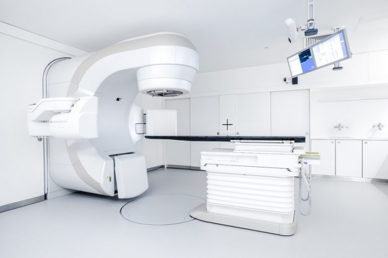
Your radiation treatments are administrated by a trained therapist in a dedicated room. The treatment is given by a linear accelerator (pictured below) while you lie on a couch. The arrangement is quite “roomy” unlike that of a cat scan or MRI. The actual treatment is painless. When receiving the treatment, you are monitored by closed circuit TV and you may converse with the staff by intercom.
While Intensity Modulated Radiation Therapy (IMRT) is very accurate, it does have a short coming. The entire plan is based upon your initial Cat Scan image which fixes the position of the internal organs. However, the prostate, bladder, rectum and other organs display some movement and there can be a variation in position of the organs relative to the initial Cat Scan of several millimeters to as much as a centimeter or two (1 cm ~ 4/10 inch).
To counter this organ movement, we acquire your image on a frequent basis (often daily). You are placed on the treatment couch in the treatment room in the usual way. However, just prior to beginning your treatment, an image is obtained to determine the relative positions of the internal organs.

